
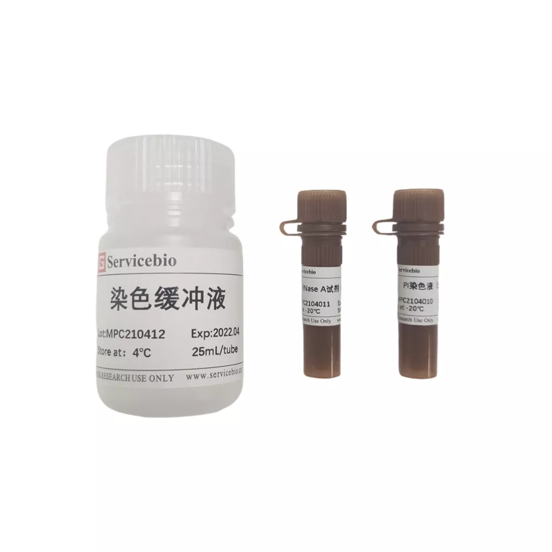
G1700-50T Cell Cycle and Apoptosis Detection Kit
The cell cycle refers to the whole process that a cell goes through from the completion of one division to the end of the next division. It is mainly divided into two phases: the interphase and the mitosis (M phase); the intercellular phase is mainly composed of the prophase of DNA synthesis (G1 phase). ), DNA synthesis phase (S phase) and late DNA synthesis (G2 phase). The sequence of changes in the entire cell cycle can be represented by G1→S→G2→M. First, the G1 period: the cell mainly synthesizes RNA and protein and other substances to prepare the cell for material and energy to enter the S phase; then enters the S phase: the cell begins to synthesize DNA and some histones, and the cell's DNA content begins to increase; finally To G2 stage: At this time, the DNA content of the cell has become twice that of the G1 period, and DNA replication has stopped, and a large amount of protein and other substances are synthesized to enter the mitosis period; if G0 (cells temporarily stop dividing and differentiation period) , Quiescent phase)/G1 phase, the DNA content in the cell is 1N; then the DNA content in the cell in the G2 phase is 2N; and the S phase cell in the G1 and G2 phase, the DNA content is between 1N and 2N; and In apoptotic cells, the nucleus will undergo condensation and DNA fragmentation, resulting in the loss of some genomic DNA fragments, so its DNA content is less than 1N. The so-called sub-G1 peak appears on the fluorescence image of flow cytometry, that is, apoptotic cells. peak. Therefore, the cycle and state of the cell can be judged according to the content of cell DNA.
Apoptosis can also be detected by observing the changes in light scattering of cells with a flow cytometer. When a cell undergoes apoptosis, apoptotic bodies are produced due to the condensation of cytoplasm and chromatin and nuclear fragmentation, which changes the light scattering properties of the cell. In the early stage of apoptosis, the chromatin shrinks, the cell density increases, and the forward angle light scattering color is significantly reduced; in the late stage of apoptosis, the cells produce apoptotic bodies, and the forward angle light scattering and lateral angle light scattering are significantly reduced.
The Cell Cycle and Apoptosis Analysis Kit uses the classic Propidium staining method to detect and analyze the cell cycle and apoptosis. Using propidium iodide can be embedded in double-stranded DNA and make it fluorescent, and the fluorescence intensity is proportional to the content of double-stranded DNA; combined with the regular changes in DNA content in different cell cycles, it can distinguish the cell cycle And status. This kit can be used for cell cycle and apoptosis detection of tissue cells, adherent or suspended cells (if it is used for tissue cell cycle and apoptosis detection, the tissue must be digested into a single cell state before the detection can be performed).
Quantity
Share :


Medical
Therapeutic Equipment
- HIFU
- Humidifier
- Ventilators
General Hospital Equipment
Diagnostic Equipment
Surgical Consumables
CareX™ robotics bedside care solutions

In Vitro Diagnostics and Life Sciences
Consumables
- PCR Consumables
- Tubes
- Micro-centrifuge Tube
- Low Retention Centrifuge Tube
- Large Volume Centrifuge Tube, Sterile
- Centrifuge Tube 5mL, Sterile
- Test Tube
- Tube Rack
- Coolbox
- Screw Cap Tube
- Screw Cap
- Transport Tube
- Cryoware
- Filtration
- Vacuum Filtration System
- Syringe Filter
- Membrane Filter
- Filter Paper
- Disposable Bottle Top Vacuum Filter
- Cell Culture & Microbiology
- Cell Culture Dish
- Cell Culture Flask
- Erlenmeyer Flask
- Cell Culture Plate
- Cell Culture Insert
- Bio-reaction Tubes
- Cell Scraper & Lifter
- Glass Coverslip
- Confocal Dish
- Chamber Slide
- Cell Strainer
- Confocal Plate
- Petri Dish
- Pipette Tips
Reagent
- Culture Media
- Buffer
- Powder
- Molecular Assay
- Nucleic Acid Preservation & Remover
- Nucleic Acid Extraction
- Reverse Transcription
- PCR Amplification
- Nucleic Acid Electrophoresis
- Protein Assay
- Protein Extraction
- Gel Preparation
- Protein Electrophoresis
- Protein Transfer
- Antibody
- ECL Kit
- Cell Separation and Digestion
- Nucleus Fluorescence Detection
- Cell Apoptosis Detection
- Cell Proliferation Detection
- Pathological
- Fixative & Dewaxing Liquid
- Staining Solution
- Antigen-Retrieval Solution
- Immunohistochemistry Kit
- Immunofluorescence Staining Kit
- Mounting Media
Monitoring Equipment

Dental
Digital 3D Solutions
Radiography & Imaging Systems
Dental Units and Accessories
Scaling and Polishing
Files and Burs
Implants Surgery
- BC and BV Series
- Implant System
- Surgical Kits
Endodontics
Maintenance Disinfection
- Sterilization Line
- Sealing Machine
General Dental Products
- Curing Light
- Distilled Water Machine
- Furniture and Designs
- Base Plate Wax
- Cotton roll dispensers
- Disposable Saliva Ejectors
- Micro Applicator
- Mixing Bowl and Spatula
- Bur holder box
- Dam Kit
- Safety Glasses
- Sectional Contoured Matrices Kit
- Disposable Dental Air Water Syringe Tips
- Denture Box
- Others
- Retractor
- Implant Tray
- Mobile Side Cabinet in Consulting Room
VetCare Solutions
Anesthesia
Dental Equipment
Critical Care Equipment
Surgical Equipment
Laboratory Diagnostics
Examination Diagnostics
Grooming Equipment
Cages
Eco Friendly Products
Consumables
SGVET Suntec
Quest Asia Technologies
SAMSUNG Ultrasound
Humanoid Robot for Elderly Care
Embodied-Intelligence Humanoid Robot for Elderly Care
- Model
Products
Media Center
About Us
© 2026 AJJ Healthcare Management Pte. Ltd
Powered by.png) Xsosys Technology (S) Pte. Ltd.
Xsosys Technology (S) Pte. Ltd.
